Nederlands
 Français
 Deutsch


 English
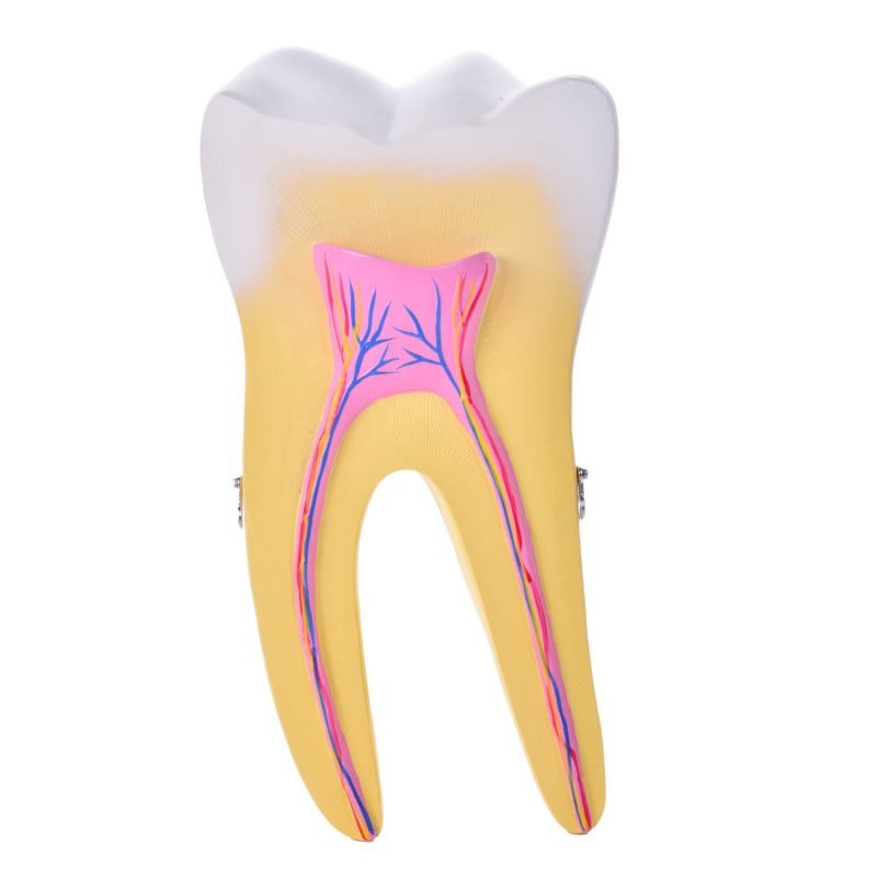
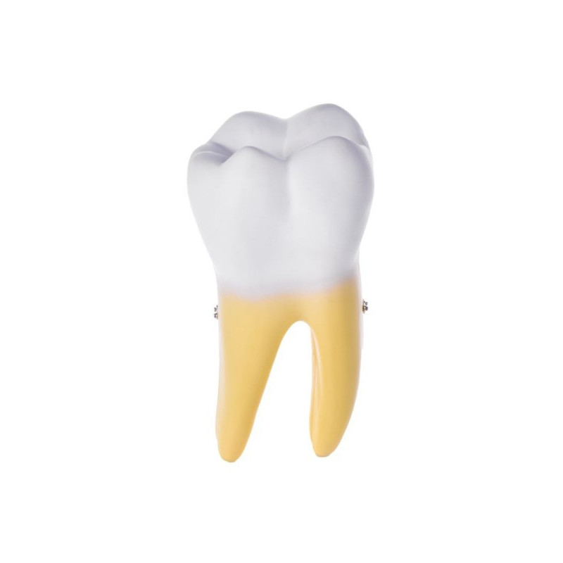
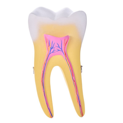
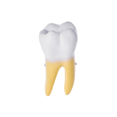
Two-Piece Molar Model – Dental Anatomy Model,This detailed two-piece molar model features a clean cross-section ideal for anatomy lessons, patient explanations and professional dental presentations.

Insured shipping

Personal support

Professional quality since 2010
Explore the internal world of a human molar with this two-piece anatomical tooth model. The model opens to reveal roots, pulp chamber, blood vessels, nerves and surrounding tooth tissues, making it ideal for anyone interested in dentistry, oral health or patient education. ?✨
Whether you are a dental student, practicing dentist or simply curious about how your own teeth are built, this model helps translate complex dental anatomy into a clear, visual explanation.
The lower molar is reproduced with a longitudinal section, so you can see the tooth from crown to root. Important structures such as enamel, dentine, pulp chamber, root canal and tooth pulp are clearly distinguishable, making this the perfect visual aid for explaining treatments and conditions.
This model is a powerful tool for lectures, chairside explanations and demonstrations. By viewing the molar from different angles, students and patients gain a deeper understanding of how tooth structure relates to decay, root canal therapy and other dental procedures.
With this molar anatomy model, you can clearly demonstrate the complex structures inside a tooth – an essential visual tool for dental schools, clinics and anyone teaching oral health.
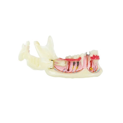
Anatomical Jaw Model – Dental Teaching Tool,This realistic anatomical jaw model opens easily and supports clear patient education, dental training and professional clinic demonstrations.
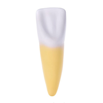
Incisor Anatomy Model – Educational Tooth Model,This realistic incisor model provides durable educational value for dental students, patient demonstrations or decorative clinic displays.